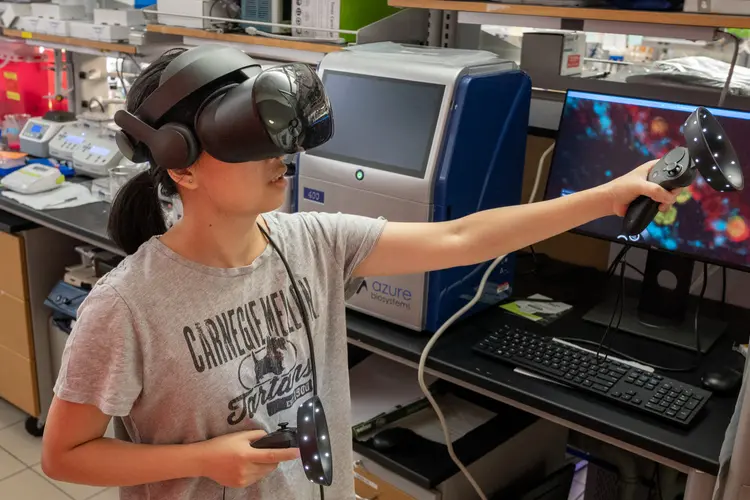
Team Brings Lung Cancer Into Focus with 3D Imaging Innovation
Media Inquiries
A new National Institutes of Health-funded collaboration at Carnegie Mellon University is breaking down barriers to high-resolution, 3D imaging of tissues — technology that could revolutionize how doctors detect, diagnose and understand cancers like lung cancer. Leon Zhao(opens in new window) of the Mellon College of Science is working with experts from the University of Illinois Urbana-Champaign and the University of Pittsburgh School of Medicine to accelerate the path from lab discoveries to clinical tools.
Finding new treatments for lung cancer requires seeing the full picture. Studying a tumor under a microscope is often like trying to understand a bustling city by looking at a single, flat photograph of one street — It’s easy to miss the critical 3D architecture and the complex interactions that define the entire system.
“Tumors are not homogenous tissue. They consist of different kinds of cells, and only some have been hijacked by the cancer,” said Zhao, Eberly Family Associate Professor of Biological Sciences. “To develop effective new strategies, there’s a strong need to understand how a tumor’s 3D microenvironment affects treatments. Our goal is to create a complete map of that environment at nanoscale resolution.”
Expanding cells for science
The Zhao Biophotonics Lab(opens in new window) is a leader in the field of enabling super-resolution imaging of biological samples through physically expanding samples in a process known as expansion microscopy.
Through the process, samples are embedded in a swellable hydrogel that homogeneously expands to increase the distance between molecules, allowing them to be observed in greater resolution.
Zhao’s Magnify technology is a variant of expansion microscopy that allows researchers to use a new hydrogel formula — also invented by Zhao's team — that retains a spectrum of biomolecules, offers a broader application to a variety of tissues, and increases the expansion rate up to 11 times linearly or about 1,300-fold of the original volume.
Zhao’s protocols have reduced the time required for sample preparation and imaging, making expansion microscopy more practical for routine use in labs and allowing nanoscale biological structures that previously only could be viewed using expensive high-resolution imaging techniques to be seen with standard microscopy tools.
“We are generating tools for cancer researchers that remove barriers so they can focus on the cancer question they want to ask,” he said.
The technique is used to precisely map structures and molecular parts of complex biological systems. For example, Zhao is producing a detailed, high-resolution map of optic nerve fibers to help make whole eye transplants a reality. He and his team also are working on nanoscale visualizations of fungi, bacteria and tumors infecting organs.
The protocol for Magnify is open source and available to the public(opens in new window). By making this powerful tool accessible and easy to use, Zhao’s lab is not only accelerating biomedical discovery but also reinforcing CMU’s commitment to democratizing cutting-edge research tools for the broader scientific community.
Discovering lung cancer’s defenses
Dr. Hua Zhang, a researcher in the Division of Malignant Hematology and Medical Oncology at University of Pittsburgh School of Medicine and UPMC Hillman Cancer Center, said that the approach the team is working on not only pushes the boundaries of high-resolution imaging but deepens understanding of how lung cancer functions at the subcellular level.
For example, the microscopy tools will open up new avenues for his research on immunotherapies for treating lung cancer. Zhang is interested in understanding how cancer cells evolve and change, particularly during the early stages of lung cancer, to allow them to evade immune detection.
“This collaboration serves as a powerful example of interdisciplinary innovation at the intersection of biomedical engineering and cancer biology,” Zhang said.
Optimized optics
Yang Liu, a professor at Illinois, specializes in instrumentation. Her lab builds custom microscopes known as omni-mesoscopes, which have large fields of view that can cover several millimeters at a time at a submicron resolution. That would be like taking a picture of a basketball court with an orbiting satellite and being able to count how many people were playing on it.
“Part of our inspiration came from astronomy. Astronomers are trying to map as many stars and objects as possible,” Liu said. “For microscopy a lot of the optics and hardware share similarities to their equipment.”
Liu, working with Hongqiang Ma, a research assistant professor in her lab, uses large-format lenses and cameras that expand the viewable area coupled with our innovative computational imaging methods to allow 3D high-resolution images to be captured. Liu also is developing an automated system to conduct the work needed to expand the cells through Zhao’s Magnify process and perform the imaging. She said that with the introduction of low-cost large-format cameras, advancements in hardware and the recent development of expansion microscopy, the timing is right for work of this magnitude.
“That’s the beauty of expansion microscopy. You’re physically expanding the samples, and the tissue is cleared with distance between its components, so it’s perfect for optical imaging,” Liu said. “By combining these technologies, we’re now able to address some of the important challenges we have had for years.”


